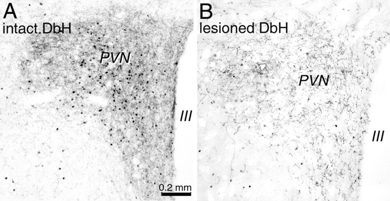
Figure 7.
Photomicrographs depicting CCK-induced cFos expression (black nuclear label) within the PVN in a nonlesioned rat (intact; A) and in a rat with a toxin-induced lesion of DbH-positive neurons (lesioned; B). Tissue sections are double labeled for DbH; note the reduced PVN cFos expression and DbH fiber labeling in the lesioned rat (B). Scale bar in A applies also to B. III, Third ventricle.

