
Figure 6.
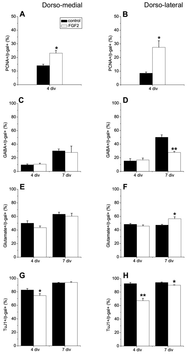
FGF2 does not induce interneurons in explant cultures. Progenitor cells from E11.5 dorsomedial (DM) and dorsolateral (DL) telencephalic walls were infected with a control retrovirus, cultured as explants in the absence or presence of FGF2 (10 ng/ml) for 3 d (4 div total), and the proportions of proliferating cells, interneurons, projection neurons, and total neurons were determined (4 div). Explants were also dissociated and cultured for an additional 3 d in monolayer cultures in the absence of FGF2 (7 div total). FGF2 did not induce interneurons in DM telencephalic explants (C) and decreased interneuron development in DL telencephalic explants after 7 div (D). Neuronal differentiation was reduced in both DM and DL explant cultures at 4 div after treatment with FGF2 (G, H), probably because of increased proliferation in these cultures (A, B). A small decrease in the percentage of neurons was maintained in FGF2-treated DL cells after 7 div, but not in DM cells (H, G). FGF2 promoted development of glutamatergic neurons in DL, but not in DM cultures (E, F). *p < 0.05; **p ≤ 0.005.