
Figure 5.
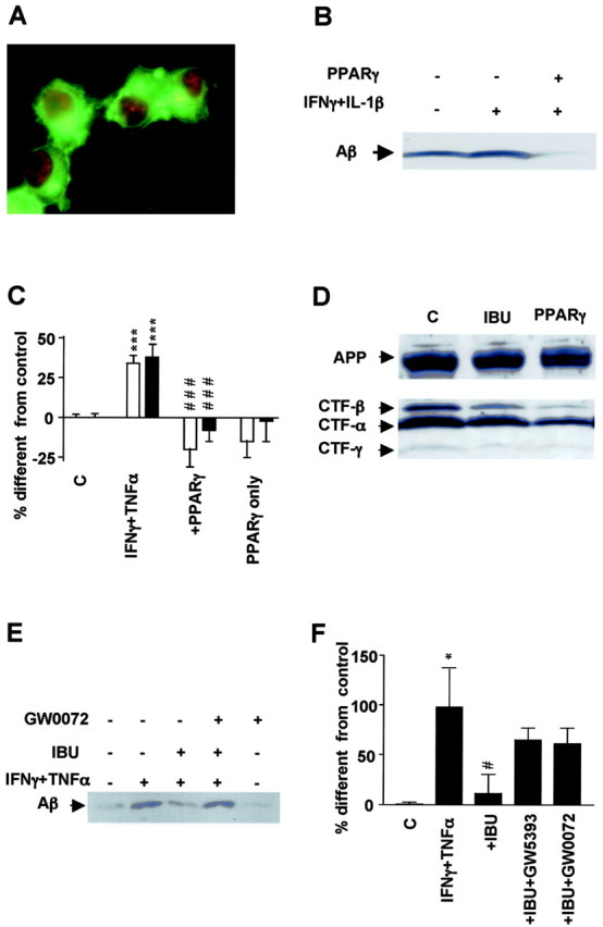
Effect of PPARγ on APP processing and Aβ secretion. A, Immunofluorescence detection of APP with 5313 antibody (green) and PPARγ with E-8 (red) in N2a cells overexpressing both proteins. B, Aβ detection by immunoprecipitation and Western blotting from media obtained from N2a cells overexpressing APPsw with or without transient transfection with PPARγ cDNA. PPARγ expression decreases generation of Aβ in cells stimulated overnight with the proinflammatory cytokines IFNγ (1 ng/ml) plus IL-1β (10 ng/ml). C, Analysis of Aβ1–40 and Aβ1–42 secretion by ELISA under the same conditions as above but in cells immunostimulated with IFNγ (1 ng/ml) plus TNFα (30 ng/ml). D, Detection of different CTFs of APP in N2a cells overexpressing APPsw and cotransfected with vector or PPARγ cDNA or incubated with ibuprofen (IBU). E, Total Aβ detection in N2A cells stimulated overnight with IFNγ plus TNFα and afterward with IBU (1μm) with or without PPARγ antagonist GW420072X (1μm) for 4 hr. F, Quantification of Aβ levels in eight experiments in which incubation with PPARγ antagonists GW0072X (1 μm) and GW5393X (1 μm) reversed the suppressive effect of ibuprofen (IBU) on APP processing. Columns represent average ± SEM. Asterisks indicate significant differences between control and treatment. Number signs represent differences between cytokines and NSAIDs. *p ≤ 0.05; ***p ≤ 0.001; #p ≤ 0.05; ###p ≤ 0.001; ANOVA followed by a Tukey's post hoc test. C, Control.