
Figure 7.
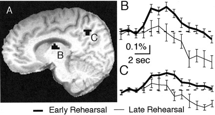
Functional MRI signal comparing early and late rehearsal. A, Activation clusters appeared in thalamus and precuneus. B, C, Time courses of activation for the left thalamus (B) and left precuneus (C). Cluster coordinates and times of significant activation are shown in Table 1.