
Figure 2.
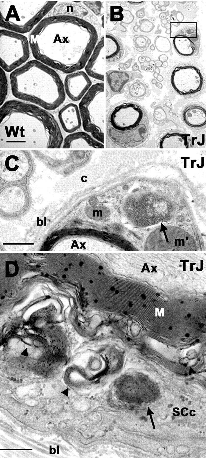
Aggresome-like structures are present in TrJ nerves. The ultrastructure of Wt (A) and TrJ (B-D) sciatic nerves are shown. A higher magnification of the area boxed in B is shown in C. Aggresome-like structures (C, D, arrows) and lamellar myelin debris (D, arrowhead) are visible in a transverse section of a TrJ nerve. Scale bars: A, B, 1 μm; C, 0.2 μm; D, 0.5 μm. Ax, Axon; bl, basal lamina; c, collagen; m, mitochondria; m′, enlarged mitochondria; M, myelin; n, nucleus; SCc, SC cytoplasm.