
Figure 4.
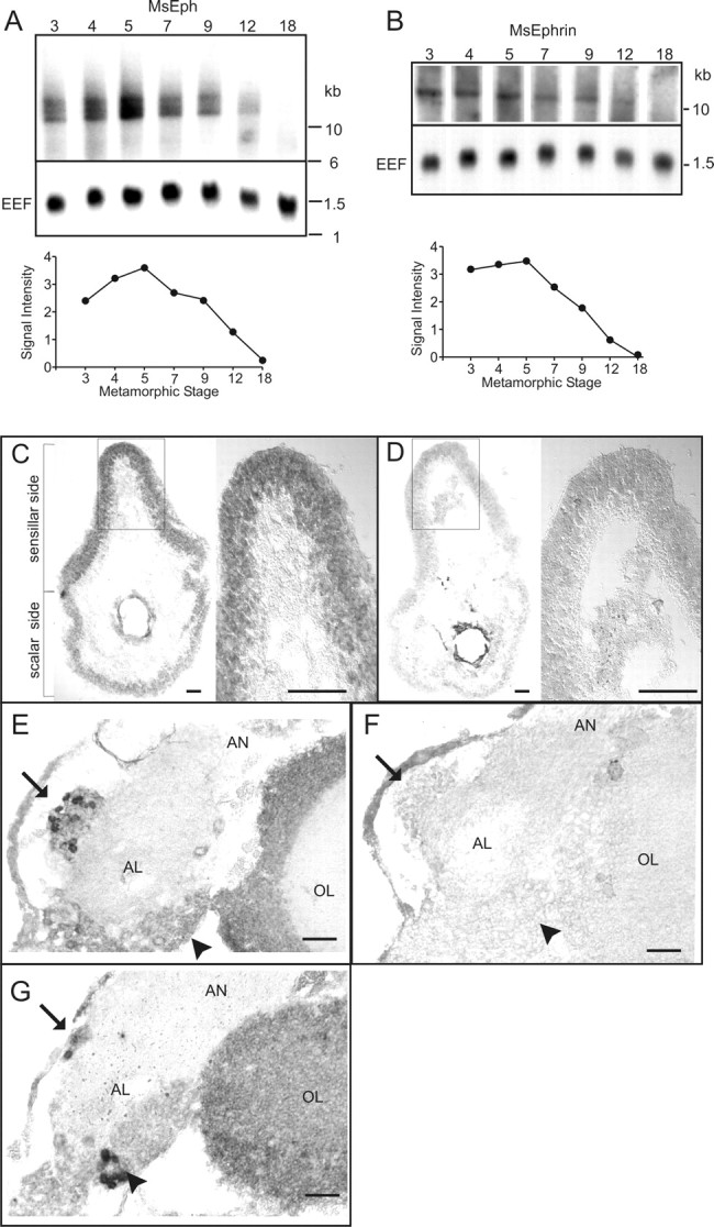
Analyses of MsEph and MsEphrin expression using Northern blots and in situ RNA hybridization. A, B, Northern blot analyses reveal developmental regulation in MsEph and MsEphrin expression in the antenna during metamorphosis. Blots hybridized with random-primed probes made against the entire MsEph or MsEphrin cDNA are shown here. Hybridization with probes made against a part of the catalytic domain of MsEph, and the core domain of MsEphrin produced indistinguishable patterns (data not shown). The Manduca EEF was used as a positive control. The graphs show relative intensities of MsEph and MsEphrin signals at each stage and are normalized to EEF signals of the same stage. C—G, Localization of the MsEph transcripts in the primary olfactory pathway by in situ RNA hybridization. Cross-sections of the antenna (C, D) and frontal sections of the AL (E-G) from stage 6 animals were hybridized with antisense probes (C, E, G) and sense probes (D, F). In C and D, left and right panels show the whole section and higher magnification images of the boxed region, respectively. In the AL, arrows and arrowheads indicate the position of the medial and lateral AL neuron packets, respectively. E-G are dorsal up and lateral to the right. AN, Antennal nerve; AL, antennal lobe; OL, optic lobe. Scale bars, 50 μm.