
Figure 2.
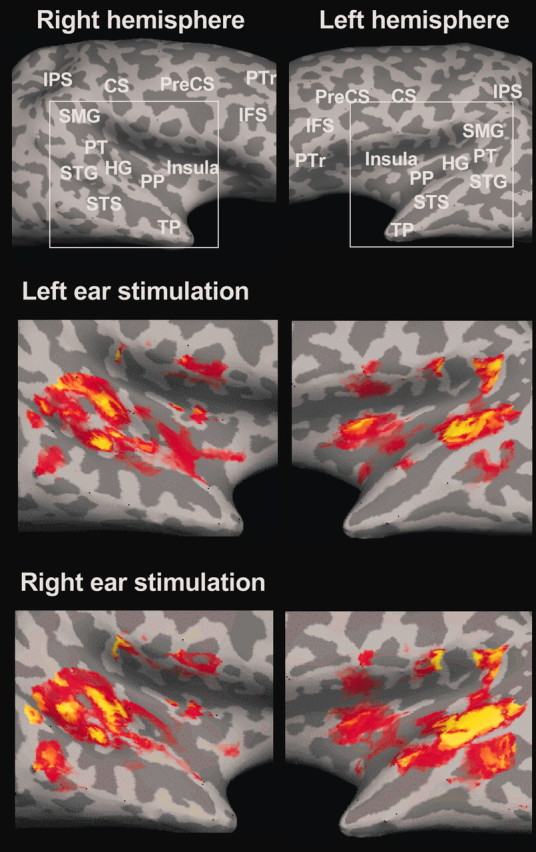
Auditory cortex activations for monaural tones relative to silence for a single participant. The top panels display lateral views of the inflated left and right hemisphere surfaces with sulci and gyri shown in dark and light gray, respectively (Fischl et al., 1999). The middle and bottom panels show activation in cortical auditory fields attributable to left and right ear stimulation. CS, Central sulcus; IFS, inferior frontal sulcus; IPS, intraparietal sulcus; PP, planum polare; PT, planum temporale; PreCS, precentral sulcus; PTr, pars triangularis; SMG, supramarginal gyrus; STG, superior temporal gyrus; STS, superior temporal sulcus; TP, temporal pole.