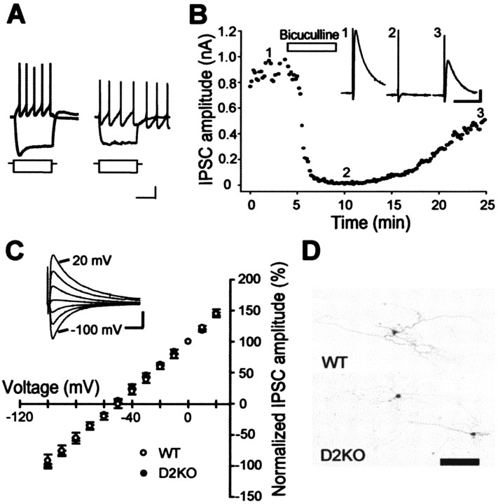
Figure 2.
Isolation of GABAA-receptor-mediated IPSCs in GP neurons. A, Superimposed voltage responses from type A (left) and B (right) neurons in response to 1000 msec hyperpolarizing and depolarizing currents. Calibration: 20 mV (voltage), 200 pA (current), 500 msec. B, Effect of bicuculline (20 μm), a GABAA receptor antagonist, on IPSCs at a holding potential of -10 mV in the presence of CNQX (10 μm) and d-AP-5 (25 μm). Traces 1-3, taken at indicated time points, are representative IPSCs in the WT mouse before, during, and after the application of bicuculline, respectively. Calibration: 500 pA, 10 msec. C, Current-voltage relationships of IPSCs recorded from D2KO (filled circles; n = 5) and WT (open circles; n = 5) mice. Current amplitudes were normalized to the value obtained at 0 mV and are expressed as means ± SEM here and in the following figures. The inset indicates representative IPSCs at each holding potential from a WT mouse. Calibration: 0.2 nA, 20 msec. D, Low-magnification micrographs of biocytin-filled GP neurons from WT (top) and D2KO (bottom) mice. Scale bar, 100 μm.

