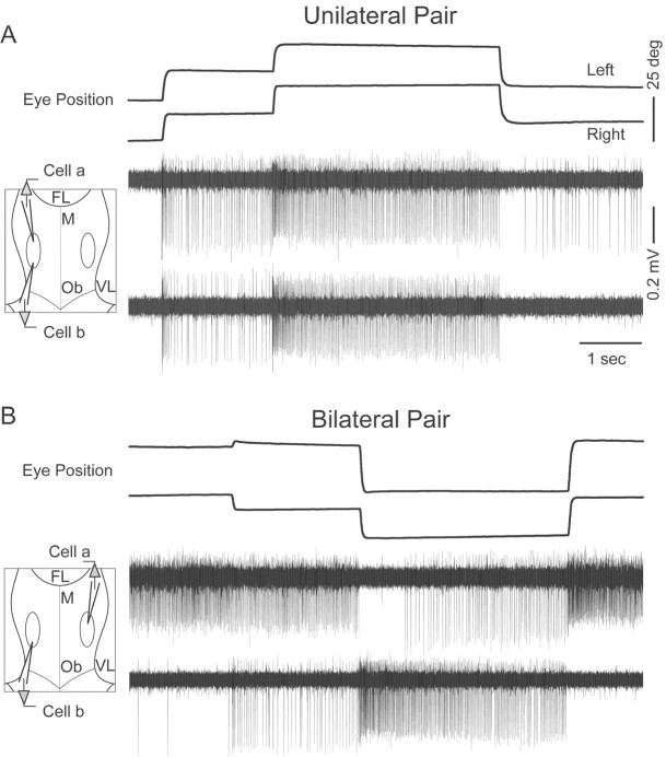
Figure 1.
Paired recordings of position cells. A, B, Eye position and extracellularly recorded potentials from position neurons on the same (A) and opposite (B) sides of midline. Insets at left are schematized views of the fourth ventricle during recording. FL, Facial lobe; M, midline; Ob, obex; VL, vagal lobe. Scale bar for B is the same as for A.

