
Figure 3.
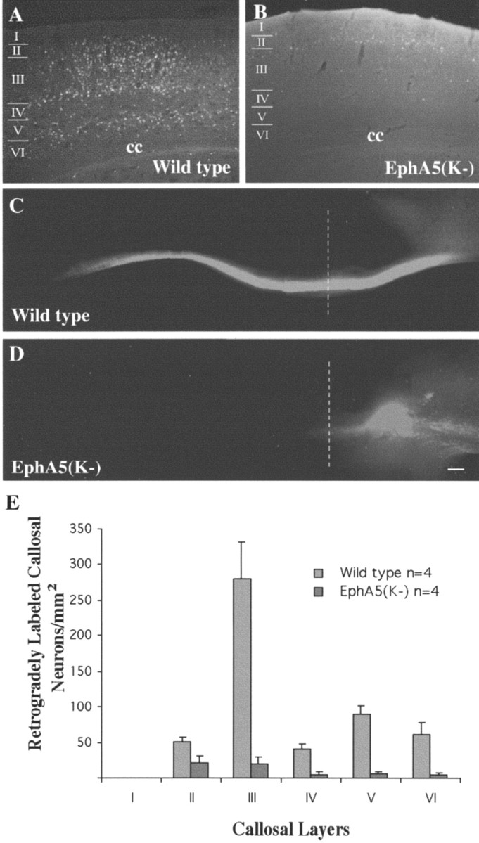
Callosal projection deficiency in EphA5(K-) adult transgenic mice. A, B, Fluoro-Gold retrograde tracing of adult wild-type (A) and transgenic (B) mice. The tracer was injected into the contralateral S1 region. Many more neurons were retrogradely labeled by Fluoro-Gold in wild type (A) than in the transgenic contralateral cortex (B). C, D, Anterograde labeling of corpus callosum with DiI in wild-type (C) and transgenic (D) mice. Dashed lines indicate the midline. E, Quantitative analysis of the distribution of callosal neurons in different cortical layers. For transgenic mice, only brains with callosal defects were analyzed. cc, Corpus callosum. Scale bar, 100 μm.