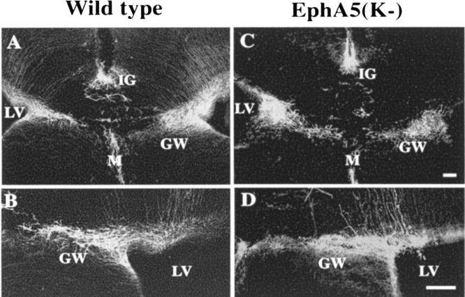
Figure 7.
Midline glia structures are normal in developing transgenic mice. A, B, GFAP-positive cells at the midline of wild-type controls. C, D, GFAP-positive cells at the midline of the transgenic mice. B and D are higher magnification photomicrographs of the glial wedge in wild-type and transgenic mice, respectively. LV, Lateral ventricle; GW, glial wedge; IG, indusium griseum; M, midline. Scale bars, 100 μm.

