
Figure 2.
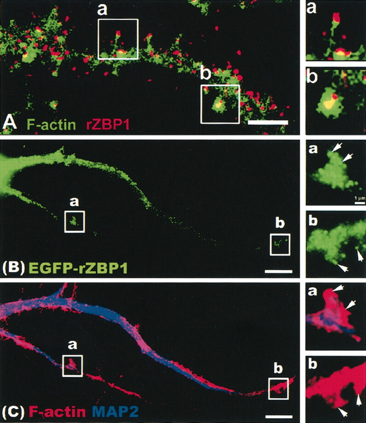
Dendritic localization of endogenous rZBP1 and EGFP-rZBP1. A, Immunofluorescence detection of ZBP1 (red) using a polyclonal antibody. F-actin was detected in the same neuron using phalloidin (Alexa 488), and images were superimposed. Two segments of interest (a,b) were enlarged (right) to show ZBP1 granules in the tip of a long, thin filopodial protrusion (a) and a spine-like, bulbous or headed protrusion (b). Scale bar, 2.5 μm. B, Transfection of EGFP-rZBP1 in cultured hippocampal neurons followed by immunofluorescence detection (C) of MAP2 (blue) and F-actin using TRITC-labeled phalloidin (red). B, C, Two dendritic segments (a,b) are enlarged (right) showing the EGFP-rZBP1 signal (B) and MAP2 and phalloidin signals (C). Arrows denote localization of EGFP-rZBP1 granules in F-actin-rich structures that protrude from the dendritic shaft. Scale bar, 10 μm.