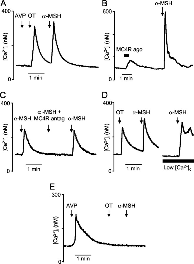
Figure 7.
Effect of α-MSH and MC4R ligands on [Ca2+]i in isolated supraoptic neurons. A, Typical example of transient increase in [Ca2+]i induced by α-MSH (1 μm) in an oxytocin neuron identified by its Ca2+ response to oxytocin (100 nm) and the absence of response to vasopressin (100 nm). B, In an oxytocin neuron, an example of a typical [Ca2+]i response to MC4R agonist compared with α-MSH response. C, Example of a neuron in which the [Ca2+]i response induced by α-MSH (100 nm) was suppressed reversibly in the presence of the MC4R antagonist (500 nm). D, Typical example showing that, in an oxytocin neuron, α-MSH (100 nm) still triggered a [Ca2+]i increase in the presence of low-Ca2+ EGTA buffer. E, Typical example of absence of [Ca2+]i response to α-MSH (1 μm) in a vasopressin neuron identified by its Ca2+ response to vasopressin (100 nm) and its absence of response to oxytocin (100 nm).

