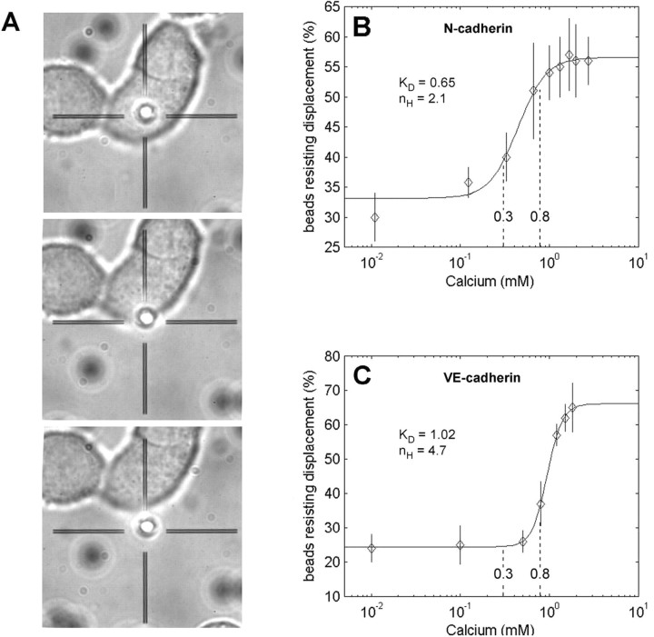
Figure 2.
Ca2+ dependency of N-cadherin and VE-cadherin binding probed by laser tweezer. A-C, Beads coated with N-cadherin-Fc (A, B) and VE-cadherin-Fc (C) settled on dorsal surface of PC12 (A, B) and MyEnd (C) monolayers were probed with laser tweezer at various extracellular Ca2+ concentrations of [Ca2+]e. A, Time series of a typical control experiment. At 0 mm [Ca2+]e, the majority of beads can be removed by laser trapping from the dorsal cell surface. B, C, Each point represents the percentage of beads (average of ≥200 beads per point) resisting displacement by laser tweezer of three to six experiments (different batches of cell cultures). Ca2+ dependency of binding of N-cadherin-Fc-coated beads displayed an apparent KD of 0.65 mm with a moderate Hill coefficient of nh = 2.1, whereas VE-cadherin-Fc-coated beads displayed an apparent KD of 1.02 mm Ca2+ and high cooperativity with a Hill coefficient of nh = 4.7. The range of the values of [Ca2+]e determined during high-frequency stimulation (0.3-0.8 mm) is indicated.

