Figure 4.
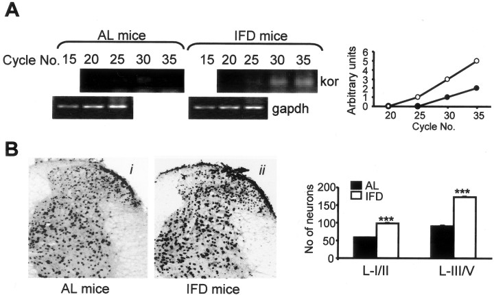
κ-Opioid receptor expression is increased in the spinal cord of IFD mice. A, Basal expression of kor mRNA in AL and IFD mice spinal cord as assessed by semiquantitative RT-PCR. gapdh mRNA served as control. Graphs represent the relative abundance of kor-specific PCR products for AL (open circles) and IFD (filled circles) mice. B, Photomicrographs and immunohistochemical analyses of the expression patterns of κ-opioid receptor in the dorsal horn of lumbar spinal cord of AL (i) and IFD (ii) mice. The histogram represents the number of positive neurons in laminas I-II and laminas III-V in each group (filled and open bars represent AL and IFD mice, respectively). We used 6-10 animals per experimental group. These animals were obtained from three independent experiments. The statistical analysis was performed by one-way ANOVA and two-tailed Student's t tests. Asterisks indicates statistical significance of the same treatments in groups AL and IFD. ***p ≤ 0.001.

