
Figure 6.
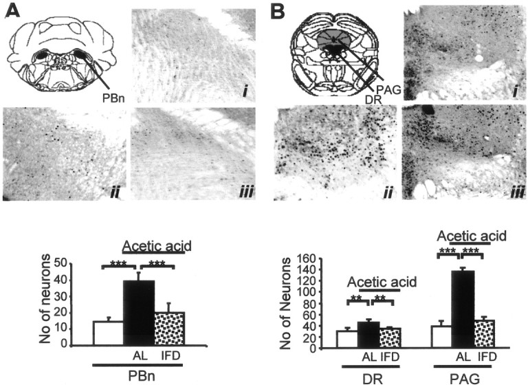
Absence of neuronal activation after noxious stimulation in brainstem nuclei of IFD mice. A, B, Photomicrographs and immunohistochemical analyses of the expression patterns of c-Fos protein in the parabrachial nucleus (A; PBn, black), a relay station of the ascending pain pathway, and in the dorsal raphe (B; DR, black) and gray periaquedultal substance (PAG; gray), components of the descending pain (modulatory) pathway of AL and IFD mice, 90 min after nociceptive treatment (ii is for AL, and iii is for IFD mice) or nontreatment (i). Histograms represent the number of positive neurons in each group. Because nontreated AL and IFD mice showed equivalent c-Fos-positive neurons, both groups are considered here as the same group (open bars); black bars are for AL animals, and dotted bars are for IFD animals treated with acetic acid. A total of five animals per group, obtained from two independent experiments, were used. The statistical analysis was performed by one-way ANOVA and two-tailed Student's t tests. *p ≤ 0.05; **p ≤ 0.01; ***p ≤ 0.001.