
Figure 7.
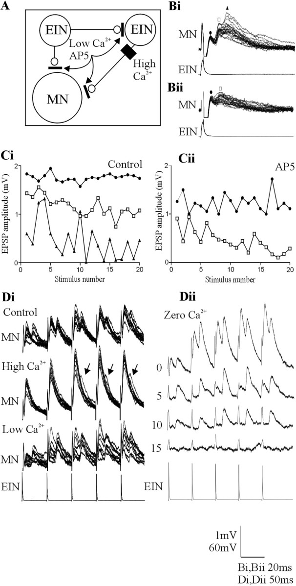
A, Summary of the proposed circuitry underlying the direct and delayed EPSPs and the methods used to examine the influence of the delayed EPSPs. Bi, An example of a triphasic feedforward EPSP in motor neurons evoked by stimulation of a single EIN. Bii, The triphasic, but not the biphasic, component was blocked by the NMDA receptor antagonist AP-5 (100 μm). The symbols above the traces refer to the graphs in Ci and Cii, which show the change in the amplitude of the initial (circle), the biphasic (square), and the triphasic (triangle) excitatory input in response to 20 Hz stimulation of the presynaptic EIN in control (Ci) and in AP-5 (Cii). Note that the triphasic input was blocked. Di, The effect of changing Ringer's solution calcium levels on the delayed EPSP. High-calcium Ringer's solution increased the amplitude of the initial EPSP. It also reduced the reliability of the delayed EPSP but did not abolish it (arrows). Low-calcium Ringer's solution reduced the initial EPSP amplitude but again did not abolish the delayed input. Dii, Traces showing the effect of zero-calcium Ringer's solution over time. Note that the monosynaptic EPSP was abolished before the delayed EPSP. The numbers at the side of the traces shows the time in minutes after application of zero-calcium Ringer's solution. MN, Motor neuron.