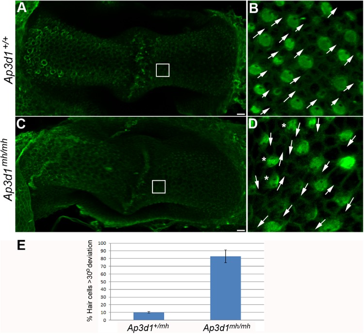
FIGURE 7:
PCP defects in the posterior crista from AP-3 mutant mice. Posterior crista from P0 control (A, B) and Ap3d1mh/mh (C, D) littermates were analyzed for hair cell orientation by staining with α-spectrin to visualize the position of the kinocilium (green). In Ap3d1mh/mh posterior crista, uniform hair cell orientation was disrupted and hair cells point in different directions (D, arrows) compared with the littermate control (B). The asterisks in D mark cells with spotty apical α-spectrin staining. Scale bars: 10 μm. (E) The misorientation of hair cells was quantified and plotted as described in Materials and Methods.

