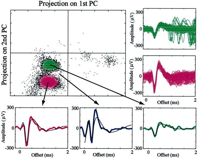
Figure 1.
Spike sorting of spikes from two neurons recorded on a single electrode. An example of off-line analysis of two cells recorded on the same electrode (990513s2e2) is shown. In the single spike examples (bottom), the actual spike shapes are depicted by the colors of the clusters, and principal-component reconstructions are plotted in black. The unidentified signal, composed of a green spike immediately followed (and distorted) by a purple spike, is shown in blue. In addition, the spike shapes of all of the spikes within the polygons are plotted (right).

