
Figure 5.
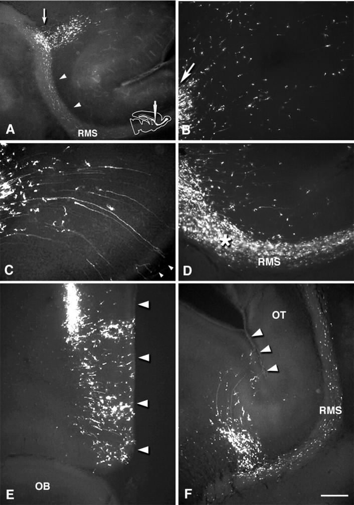
Distribution of GFP-labeled cells in vibratome sections 1 and 3 d after rostral injection. Medial sagittal section at the septal nuclei level (A–D), a coronal section at the injection level (E), and a horizontal section containing the RMS (F). A, 1 dpi. Within the SVZ, labeled cells are distributed rostrally from the injection site (arrow) into the RMS (arrowheads), and cells also migrate radially into the overlying white matter and cortex. B, C, 3 dpi. High-power field view of labeled cells in the frontal white matter and cortex. Cells migrate in a radial manner fanning out toward the pia. Arrow in B indicates labeled cells migrating out of the injection site. In the inferior frontal cortex, cells show a ventrally skewed migration (C). Some cells have long radial processes, which reach the pial surface (C, arrowheads). D, High-power field view of the RMS. Labeled cells migrate out of the vertical limb of the RMS in a direction perpendicular to the axis of the RMS. Asterisk indicates the bend between the vertical and horizontal limbs of the RMS. From coronal (E) and sagittal (F) views, it is clear that the cells migrate rostromedially toward the frontal cortex. Arrowheads indicate the medial edge of the frontal lobe. RMS, Rostral migratory stream; OT, olfactory tract. Scale bar: A, 400μm; B–F, 200 μm.