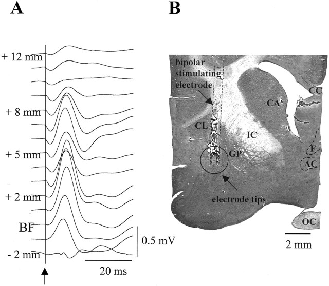
Figure 1.
A, Field potentials recorded in S1 evoked by electrical stimulation through a bipolar electrode while it was advanced from the cortical surface toward the BF. See Materials and Methods for details. The arrow and the vertical line indicate the beginning of the electrical stimulation. B, Histological localization of BF stimulation sites. The coronal section shows a lesion made trough the stimulation electrode. Vertical dotted lines mark the track of the bipolar electrode. The open circled area signals the location of the electrode tips. AC, Anterior commissure; CA, caudate; CC, corpus callosum; CL, claustrum; F, fornix; GP, globus pallidus; IC, internal capsule; OC, optic chiasm.

