
Figure 2.
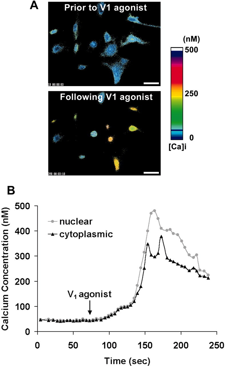
Cytoplasmic [Ca2+]c and nuclear [Ca2+]n rise in response to V1 agonist in cortical astrocytes. Cortical astrocytes were loaded with fura-2, and Ca2+ images were recorded with the InCyt2 fluorescence imaging system. A, Fura-2-generated Ca2+ images under basal conditions and after addition of V1 agonist for 100 sec. V1 agonist induced a marked nuclear Ca2+ compartmentalization. Scale bar, 30 μm. B, Window apertures were used to locate the cytoplasm and nucleus of one representative astrocyte, and [Ca2+]c and [Ca2+]n were plotted against time. V1 agonist was added where indicated and was present throughout the entire observation period. Note that Ca2+ was increased initially in both the cytoplasm and nucleus, followed by a rapid and transient Ca2+ localization into the nucleus. After the rise in [Ca2+]n, translocation of nuclear Ca2+ back to the cytoplasm occur red before there turn to baseline total [Ca2+]i.