
Figure 6.
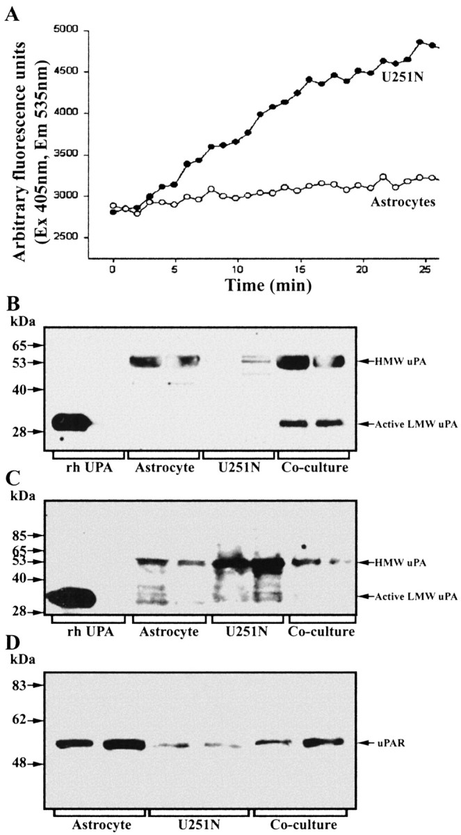
Contribution of components of the uPAR–plasmin cascade by astrocytes or glioma cells. A, Plasminogen content was determined by the use of the fluorochrome-conjugated plasmin substrate AFC-80; here, 10 μM uPA was applied to conditioned media to convert plasminogen to plasmin, the activity of which was monitored. Note that the U251N glioma cells (open circles), rather than astrocytes (filled circles), were the principal sources of plasminogen. This result was replicated three times. B, C, Western blots of cell lysates or conditioned media, respectively, of uPA expression by astrocytes and U251N cells. The cell lysate analyses (B) show high uPA levels for astrocytes, but not for U251N cells, whereas the converse is true for conditioned media (C). D, Western blot for uPAR that documents higher expression in astrocytes rather than in glioma cells.