
Figure 7.
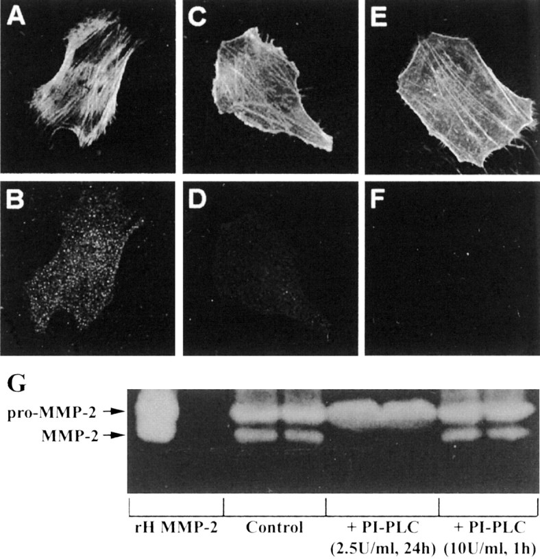
Detection of uPAR on astrocytes by immunofluorescence. Astrocytes have high expression of uPAR (B), whereas U251N cells have very low levels (D). Astrocytes (A) and glioma cells (C) were counterstained with phalloidin, which binds to filamentous actin, to delineate the boundary of each cell type. The specificity of the uPAR antibody was tested with an uPAR peptide (Santa Cruz Biotechnology, Santa Cruz, CA); when the uPAR antibody was preincubated with the uPAR peptide, subsequent staining of astrocytes with this antibody mixture was negative (data not shown). E, F, Astrocytes were treated with PI-PLC (2.5 U/ml) for 24 hr and then immunostained for uPAR. After PI-PLC treatment the astrocytes (E; phalloidin staining) were no longer immunoreactive for uPAR (F). Finally, continuous 2.5 U/ml PI-PLC for 24 hr (but not 10 U/ml PI-PLC for 1 hr, followed by 23 hr of recovery) abrogated the activation of MMP-2 in glioma–astrocyte interaction (G).