Figure 1.
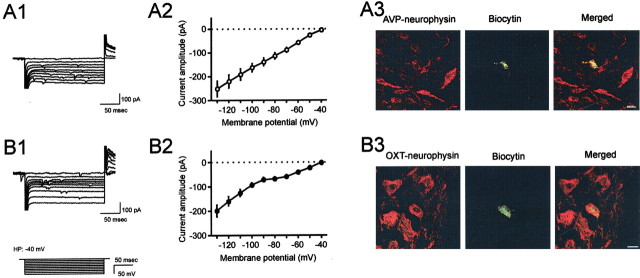
The presence or the absence of voltage-activated membrane currents characterize two distinct subpopulations of magnocellular neurons. A1, Superimposed current traces representative of a subpopulation of magnocellular neurons showing a linear I-V relationship in response to 200 msec hyperpolarizing voltage steps elicited from a holding potential of −40 mV. The voltage paradigm is depicted at bottom left. A2, A mean I-V plot of this population (n = 10). B1, A representative of another subpopulation of magnocellular neurons shows a different I-V relationship in response to the same protocol. B2, The mean I-V plot from 10 magnocellular neurons showing a similar nonlinear relationship. A sustained outward rectification (SOR) is observed at membrane potentials more positive than −70 mV, whereas a hyperpolarization-induced inward rectification (IR) appears at membrane potential more negative than −90 mV. A3, Confocal image of a cell that did not display SOR/IR, filled with biocytin by electroporation through perforated patch (green). This cell was colabeled with AVP-associated neurophysin antibody (red). B3, Another cell that showed SOR/IR, which was filled with biocytin (green) and was OXT-associated neurophysin immunoreactive (red). Scale bars, 10 μm.

