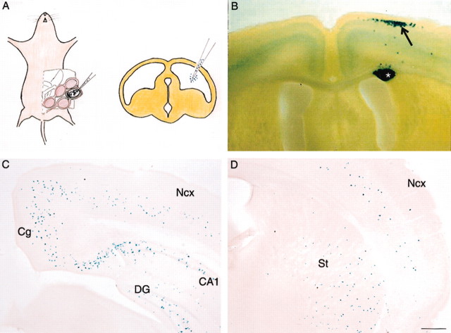
Figure 1.
In utero transplantation of βgal-labeled progenitor cells results in their incorporation into cortical and extracortical structures. A, Schematic diagram to illustrate introduction of cell suspensions into the developing ventricle using a transplacental approach. The loaded pipette is introduced into the ventricle, and cells are injected by pressure. B, Transplantation of progenitors from the neocortical VZ typically generates clumps of labeled cells (asterisk) in the white matter, with associated labeling of cortical neurons lying radially above (arrow). In this example, cells introduced into E14.5 hosts have migrated as a cluster to the upper cortical layers. C, D, Transplanted MGE cells do not form radial clusters; instead, they give rise to scattered cells in the neocortex (Ncx) and other structures, including cingulate cortex (Cg), hippocampal dentate gyrus (DG) and CA fields (CA1), and striatum (St). Scale bar (in D): B, 570 μm; C, D, 375 μm.

