
Figure 3.
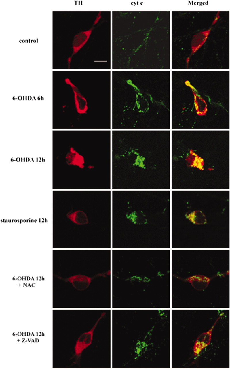
Colocalization of cytochrome c with TH-positive neurons after 6-OHDA treatment. Primary cultures of mesencephalic DA neurons were treated with 20 μm 6-OHDA or 1 μm staurosporine for the time periods indicated in the presence or absence of 50 μm zVAD-fmk (Z-VAD) or 0.5 mmN-acetylcysteine (NAC). Cultures were double immunolabeled with a rabbit polyclonal anti-TH and a mouse monoclonal anti-cytochrome c (cyt c) and were followed by incubation with Alexa Fluor 568 goat anti-rabbit IgG and Alexa Fluor 488 goat anti-mouse IgG as described in Materials and Methods. Cultures then were examined by confocal microscopy. Scale bar, 10 μm.