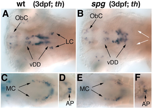
Figure 4.
The catecholaminergic neurons in the diencephalon are reduced and the LC is absent in spg mutant embryos. A–F, th expression in the forebrain and hindbrain of wild-type (A, C, D) and spg (B, E, F) mutant embryos at 3 dpf. A–F, Dorsal views, anterior is to the left. In spg mutant embryos (B), the catecholaminergic neurons of the LC are absent and those of the diencephalon are reduced, whereas the remaining hindbrain CA clusters develop normally (E, F). AP, Area postrema; vDD, ventral diencephalic dopaminergic neurons; MC, medullary catecholaminergic neurons; ObC, olfactory bulb catecholaminergic neurons; white arrows indicate the absence of the LC.

