
Figure 2.
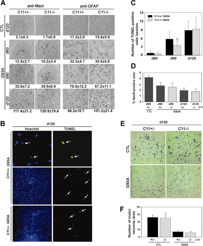
The lack of caspase-11 failed to block neurodegeneration in the G93A mice. A, Glial activation is not affected by the absence of caspase-11. The lumbar spinal cords from caspase-11 single mutant (CTL) and caspase-11; G93A double-mutant mice were taken at indicated days of age and processed for immunohistochemistry using anti-MacI antibody to detect microglial cells and anti-GFAP antibody to detect astrocytes. The cells positive for anti-MacI or anti-GFAP were counted in the white matter of the spinal cord sections, and the numbers are shown at the bottom of each panel (n = 5 for single mutants; n = 12 for double mutants; mean ± SD). Original magnification for the anti-MacI staining, 20×; anti-GFAP staining, 40×. B, TUNEL-positive apoptotic cell death in G93A mice is not significantly affected by the absence of caspase-11. Spinal cords of indicated genotypes and days of age were processed for TUNEL. Arrows indicate the TUNEL-positive cells (top panels, 63× magnification; bottom panels, 20× magnification). Note the condensed nuclei of TUNEL-positive cells in the top panels. C, The numbers of TUNEL-positive cells were determined from the lumbar sections by direct counting (n = 30). *p < 0.05 indicates significantly different (Student's t test). D, Neuronal damage–loss is not protected by the absence of caspase-11. Total neuronal area was measured by staining the spinal cord sections with a neuronal marker anti-NeuN and scanning the positive area by Northern Exposure (n = 10; mean ± SD) and was confirmed by visual inspection. E, F, Motor neuron loss is not protected by caspase-11 deficiency in G93A mice. Number of motor neurons was determined by Nissl-staining the spinal cord sections (E) and counting the large Nissl-positive neurons (F). Three 120-d-old mice were used for each genotype, and 10 tissue sections of lumbar spinal cord were counted for each mouse. Data represent the mean of pooled counting with SD.