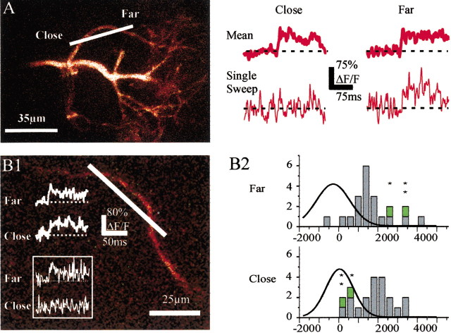
Figure 3.
Measurements of fast Ca2+ transients underestimate action potential backpropagation. A, Ca2+ signals were recorded simultaneously at two points from the same first-order branch of a tuft. Average Ca2+ transients (including failures) were similar at both locations (top traces). In some cases, however, an action potential did not trigger any Ca2+ signal at the site close to the soma whereas it did farther (bottom traces). B1, Ca2+ signals were recorded simultaneously at two close points from the same branch of a basal dendrite. Average Ca2+ transients (including failures) were similar at both locations (top traces). Occasionally, an action potential did not trigger any Ca2+ signal at the site closer to the soma whereas it did farther (bottom traces, inset). B2, Integral histograms of ΔF/F measured over 50 msec after the action potential peak at the recording sites closer to (Close) and farther from (Far) the soma on the basal dendrite shown in B1. The green symbols (and the above stars) identify two cases of apparent failures at the proximal site and the corresponding calcium transients at the distal site. The superimposed curves correspond to the Gaussian fits of the noise integral distributions.

