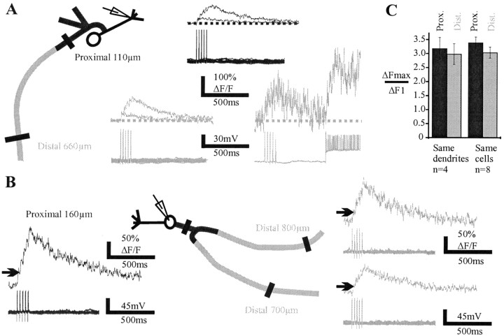
Figure 6.
Burst of action potentials backpropagate distally in basal dendrites. A, Left, Superposition of Ca2+ signals evoked by one and five action potentials (interspike interval = 25 msec) in the proximal (top traces) and distal (bottom traces) parts of abasal dendrite (average of 10–18 sweeps). The recording electrode was located near the first branching point of the apical dendrite. The summation of fluorescence was sublinear at each site. The sublinearity was not caused by saturation of the dye because the Ca2+ signal could still increase significantly during higher firing frequency, as shown for the most distal measurement. B, Another cell in which the amplitude of ΔF/F differed in two distal branches originating from the same proximal branch. In that case summation was nearly linear. C, Means of ΔFmax/ΔF1 recorded in the proximal and distal parts of basal dendrites. Histograms on the left are from four different cells in which measurements were obtained on the same dendritic branch (no branching points in between). Histograms on the right include additional recordings in which branching points were present between the proximal and distal recording sites.

