Figure 8.

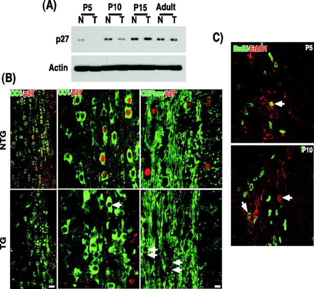
P27 level is significantly decreased in the p75–ErbB1 KD mice. A, Quantification of p27 in Western blot analyses. Actin blot was used as a control. B, The spinal cord sections taken from P10 mice were stained for CC1 and p27 or CNPase and p27. Left, Low-magnification pictures; scale bar, 20μm. Middle and right, High-magnification pictures; scale bar, 8.3μm. Note that some cells in the p75–ErbB1 KD mice have p27 in the nucleus, but its level is much lower than that found in NTG (arrows). C, The p75–ErbB1 KD is expressed among some dividing oligodendrocyte progenitors. The p75–ErbB1 KD was detected by ErbB1 staining in the cerebellar white matter at P5 and P10. Note that the cells that are positive for ErbB1 contain BrdU immunoreactivity in their nucleus.
