
Figure 6.
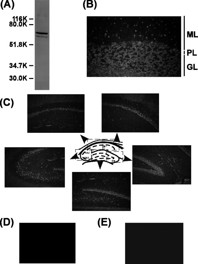
Immunohistochemical localization of Ali1 in brain. A, Total brain extract (100μg) was subjected to 10% SDS-PAGE, and Western blotting was performed with anti-rat Ali1 antiserum. B, Immunohistochemical localization of Ali1 in the rat cerebellum. ML, Molecular layer; PL, Purkinje cell layer; GL, granule cell layer. C, Immunohistochemical localization of Ali1 in the rat hippocampus. D, Negative control staining of the rat cerebellum. E, Negative control staining of the rat hippocampus.