
Figure 1.
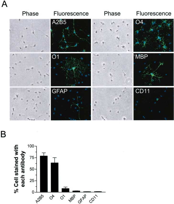
Characterization of primary OL cultures.A,Representative microphotographs of morphology and immunocytochemistry of cells (7–9 d in vitro) labeled with indicated antibodies. B, Composition of developing OL cultures as determined by immunostaining for indicated markers. Total cell number was determined by counting all cells labeled with the nuclei dye Hoechst 33258. Values represent mean ± SEM from three separate experiments.