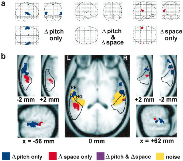
Figure 2.
Statistical parametric maps for contrasts of interest (group data). a, SPMs are shown as “glass brain” projections in sagittal, coronal, and axial planes. b, SPMs have been rendered on the group mean structural MRI brain image, normalized to the MNI standards terotactic space (Evans et al., 1993). Tilted axial sections are shown at three levels parallel to the superior temporal plane: 0 mm (center), +2 mm, and -2 mm (insets). The 95% probability boundaries for left and right human PT are outlined (black) (Westbury et al., 1999). Sagittal sections of the left (x =-56 mm) and right (x =+62 mm) cerebral hemispheres are displayed below. All voxels shown are significant at the p < 0.05 level after false discovery rate correction for multiple comparisons; clusters less than eight voxels in size have been excluded. Broadband noise (without pitch) compared with silence activates extensive bilateral superior temporal areas including medial Heschl's gyrus (HG) (b, center, yellow). In the contrasts between conditions with changing pitch and fixed pitch and between conditions with changing spatial location and fixed location, a masking procedure has been used to identify voxels activated only by pitch change (blue), only by spatial change (red), and by both types of change (magenta). The contrasts of interest activate distinct anatomical regions on the superior temporal plane. Pitch change (but not spatial location change) activates lateral HG, anterior PT, and planum polare (PP) anterior to HG, extending into superior temporal gyrus, whereas spatial change (but not pitch change) produces more restricted bilateral activation involving posterior PT. Within PT (b, axial sections), activation attributable to pitch change occurs anterolaterally, whereas activation attributable to spatial change occurs posteromedially. Only a small number of voxels within PT are activated both by pitch change and by spatial change.

