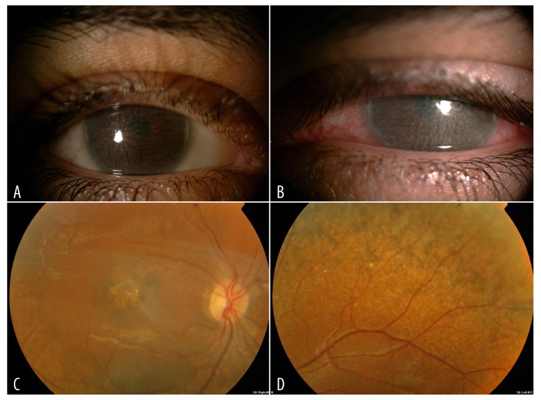
Figure 2.
(A) Follow up clinical photo in the same case with increasing corneal deposits and haze in the right eye. (B) Follow up clinical photos in the same case with increasing corneal deposits and haze in the left eye. (C) Fundoscopy image showing the right eye macular changes. (D) Fundoscopy image showing the left eye peripheral pigmentary changes.

