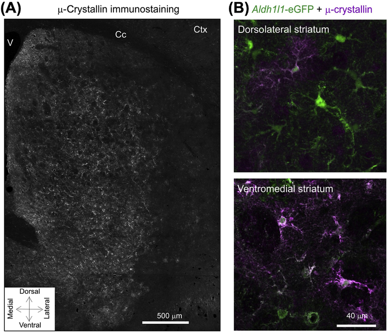
Figure 2. μ-Crystallin Displays a Gradient of Expression within Striatal Astrocytes.
(A) μ-crystallin immunostaining in striatum showing its spatial gradient. There are higher levels of expression in the ventral striatum compared with dorsal areas. (B) Representative images for μ-crystallin immunostaining in dorsolateral and ventromedial parts of the striatum in brain sections from Aldh1l1-eGFP mice. In the dorsolateral area, ~30% of astrocytes were μ-crystallin positive, whereas, in the ventromedial area, this was ~90%. Adapted from [50]. Abbreviations: Cc, corpus callosum; Ctx, cortex; V, ventricle.

