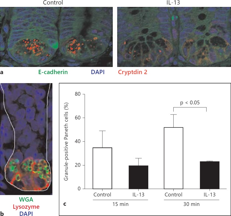
Fig. 4.
IL-13-induced Paneth cell degranulation. a Small intestinal tissue sections from C3H/HeN mice treated i.p. with 500 µl of PBS (left panel, control) or IL-13 (300 ng/animal, in 500 µl; right panel) for 30 min were stained for cryptdin 2 (red) and E-cadherin (green). Counterstained with DAPI. Original magnification ×630. b Small intestinal tissue sections were stained for lysozyme P (red) and mucus (green) to illustrate the anatomical position of granulated Paneth cells. Counterstained with DAPI. Original magnification ×630. c Quantitative determination of Paneth cell granulation (%) every 15 and 30 min after administration of PBS (control) or IL-13.

