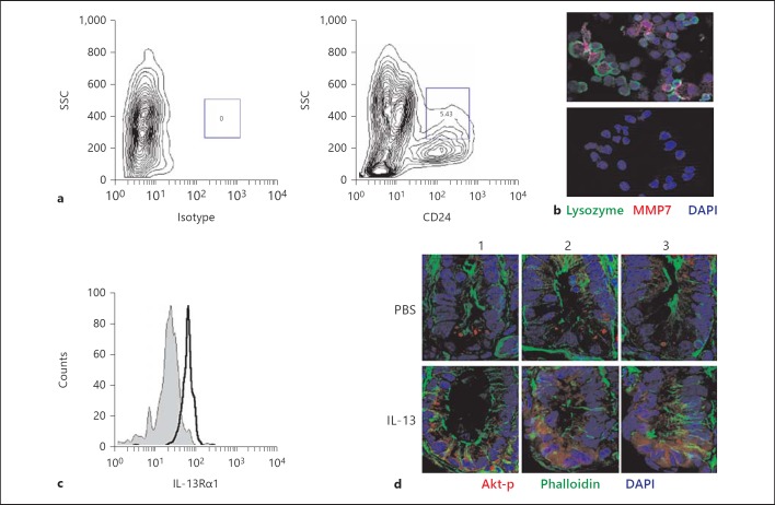
Fig. 5.
Paneth cells express the IL-13 receptor IL-13Rα1 and respond to stimulation. a Small intestinal crypts were isolated, trypsinized, stained for CD24 and analyzed by flow cytometry. Paneth cells are gated as CD24hiSSChi cells resulting in 4–6% of all CD45-E-cadherin+ cells. b CD24hi cells were sorted and immunostained for lysozyme P and MMP7 expression. The lower panel depicts staining with isotype controls. Counterstained with DAPI. Original magnification ×630. c Isolated cells were stained using an anti-IL-13Rα1 antibody or an isotype control and CD24hi cells were analyzed by flow cytometry for expression of the IL-13 receptor. d Immunohistological staining of phosphorylated Akt in sections of the small intestine of mice 30 min after injection of PBS or IL-13 (300 ng) i.p. Images of three individual crypts (1-3) visualized in small intestinal tissue sections from PBS- and IL-13-treated mice are shown. Counterstained with phalloidin (green) and DAPI (blue). Original magnification ×400.

