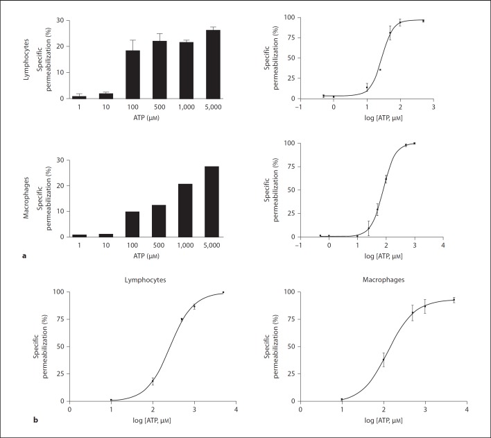
Fig. 1.
MLN and peritoneal cells were permeabilized at different ATP concentrations. Mononuclear cells from MLN (a) or the peritoneal cavity (b) were removed and adjusted to 2 × 10⁵ cells/sample. Cells were incubated with increasing concentrations of ATP for 10 min at 37°C. During the last 5 min of incubation EB was added and samples analyzed by flow cytometry. a MLN cells, individual experiments performed in triplicate, n = 15. b Peritoneal cells, individual experiments performed in triplicate, n = 7.

