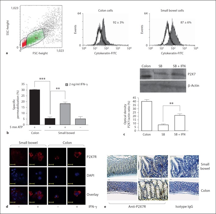
Fig. 7.
Epithelial cells from the colon but not from the small bowel permeabilize following incubation with ATP. Colonic and small bowel epithelial cells were isolated and adjusted to 2 × 10⁵ cells/sample. Epithelial cells were preexposed to 2 ng/ml IFN-γ for 24 h, incubated with 5 mM ATP for 10 min at 37°C (b) or processed for Western blot analysis (c) and confocal microscopy (d). During the last 5 min of incubation EB was added and samples analyzed by flow cytometry. ** p < 0.001, *** p < 0.0001 (a, b). a Epithelial cells were selected on dot plot gate R2 and cytokeratin-FITC staining. d Confocal microscopy of cytospin preparations showing the relative distribution and levels of P2X7 receptor (red) in freshly isolated epithelial cells from the small bowel and colon. Nuclei stained with DAPI (blue). Representative of 3 experiments from each site. Scale bars represent 20 µm. e P2X7 receptor immunostaining (brown) is shown in the small bowel and colon of normal mice. Representative of 6 independent samples. Scale bars represent 50 µm. SB = Small bowel.

