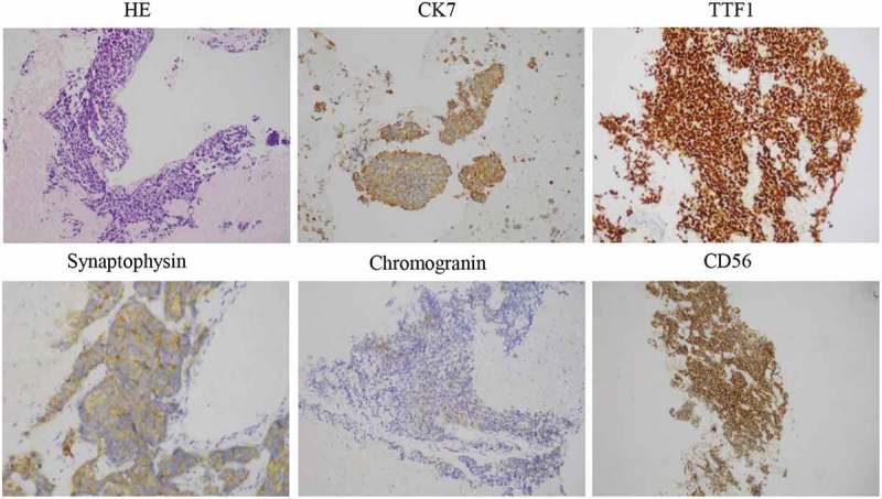
Figure 3.

HE and IHC staining was performed on primary tumor biopsies after 6 months of erlotinib treatment. The cells displayed an SCLC phenotype with hyperchromatic nuclei, abundant cytoplasm, and inconspicuous nucleoli. Typical for SCLC, IHC was strongly positive for CD56 and TTF1, and focally for CK7 and synaptophysin (all 400×).
