Fig. 1.
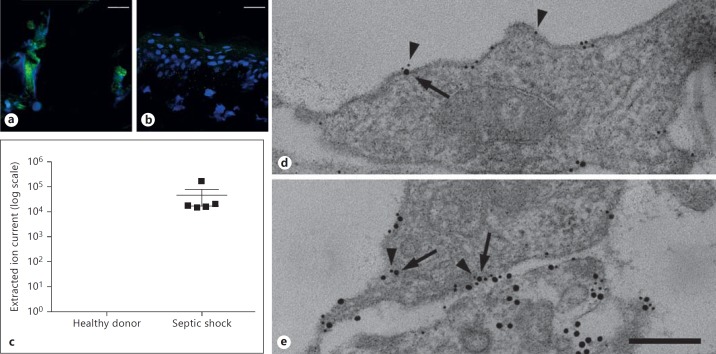
Detection of extracellular histone H4. Immunostaining of tissue from a patient with cellulitis caused by an S. pyogenes infection (a) and a healthy volunteer (b) are depicted. Histones were detected with an antibody against histone H4. Histones are stained in green and DNA (DAPI) in blue. Scale bar: 25 μm. c MS data on histone H4 release in patients (n = 5) with septic shock or healthy donors (n = 5). Detectable amounts of histone H4 are presented as extracted ion current, which describes the intensity (area under curve) of a selected precursor mass from all peptides measured. d The endogenous expression of p33 (5 nm gold) on noninfected HUVEC is shown. Note, that almost no release of histone H4 was detected. e Treatment of HUVEC cells with S. pyogenes evokes cell necrosis and subsequent release of histone H4 (10 nm gold) that colocalizes with endogenous p33 (5 nm gold); p33 (5 nm gold, arrowheads) and histone H4 (10 nm gold, arrows) were immunostained with gold-labeled antibodies. Scale bar: 100 nm.

