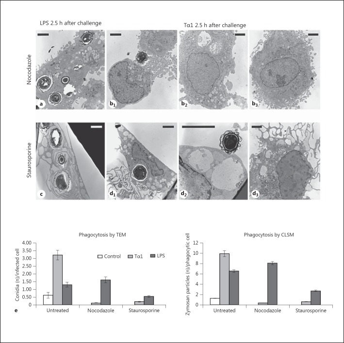
Fig. 8.
Effect of microtubule destabilization by nocodazole and of PKC inhibition by staurosporine on the phagocytic activity of Tα1-stimulated human MDMs. a-d TEM images showing the A. niger conidial uptake in LPS-stimulated (a, c) and Tα1-stimulated MDMs (b, d) in the presence of nocodazole (a, b) or staurosporine (c, d); b1, b2, b3, d1, d2, d3 Different representative cells in Tα1-stimulated MDMs. Bars = 2 µm. e Phagocytosis of A. niger conidia by Tα1- or LPS-stimulated MDMs in the absence and presence of nocodazole or staurosporine; the solid bar graphs report the number of internalized conidia evaluated by TEM (left graph) and the number of nonopsonized zymosan particles evaluated by CLSM (right graph). Significance vs. untreated control: Tα1 + nocodazole, p = 0.0156927; LPS + nocodazole, p = 0.121706; Tα1 + staurosporine, p = 0.017701; LPS + staurosporine, p = 0.037381.

