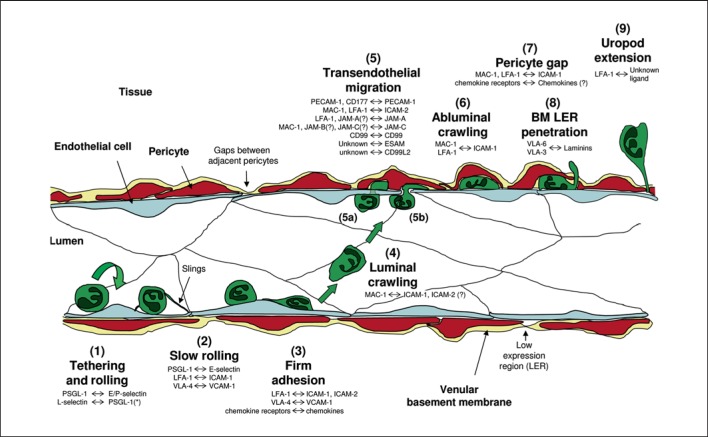
Fig. 1.
The neutrophil adhesion cascade. The diagram depicts a simplified illustration of the neutrophil adhesion cascade that mediates migration of neutrophils from the vascular lumen to the extravascular tissue. The distinct steps are indicated by numbers (1-9) and the key associated molecular pathways are listed with the molecules named on the left-hand side being neutrophil-expressed and on the right-hand side being expressed by the vessel wall (i.e. ECs, pericytes or venular BM). * The ligand of neutrophil-expressed L-selectin is neutrophil-derived PSGL-1 (i.e. neutrophil rolling on adherent neutrophils or EC-bound neutrophil particles). The initial step is (1) the capture and rolling of neutrophils on activated ECs followed by (2) slow rolling (largely mediated via selectin signalling) and (3) firm adhesion to ECs through strong adhesive contacts between leucocytes and ECs mediated through activation of neutrophil integrins by chemo-attractants such as chemokines. These interactions induce intracellular signals within the neutrophils that are responsible for cytoskeletal rearrangements and polarisation of the cell. This leads to (4) luminal motility (or luminal crawling) along the endothelium until they reach their site of (5) TEM. Neutrophils can cross the endothelium by either (5a) transcellular or (5b) transjunctional routes (the latter being more prevalent). This step involves complex interactions involving numerous EC junctional molecules. Once through the endothelium, neutrophils exhibit (6) abluminal crawling along pericyte processes. At this step, neutrophils will also be interacting with the venular BM, most probably via integrin-mediated mechanisms. Neutrophils eventually breach the pericyte layer (7) by migrating through gaps between adjacent cells and (8) regions of low matrix protein deposition (LERs) in the BM. To finally migrate into the interstitial tissue, neutrophils need to detach from the vessel wall (9), a response that is illustrated through the formation of an elongated uropod that must be detached before neutrophils can initiate their movement towards the inflammatory foci.

