Figure 5.
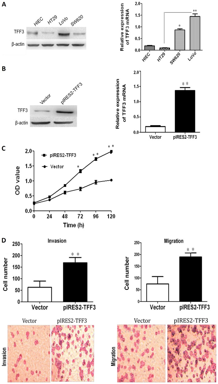
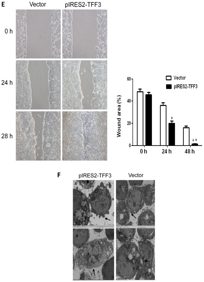
The overexpression of TFF3 promotes the proliferation, migration and invasion capacities of the HT29 cells. (A) Both western blot analysis and RT-qPCR revealed that TFF3 expression in the HT29 and LoVo cells were the lowest and highest, respetively. (B) TFF3 expression was found to be enhanced significantly at the mRNA and protein level following transfection with the TFF3 expression plasmid. (C) CCK-8 assay revealed that the overexpression of TFF3 markedly increased the proliferation of the HT29 cells. (D) Transwell chamber assay displayed that the migration and invasion of the HT29 cells was markedly enhanced following the overexpression of TFF3. (E) The migration of the HT29 cells was also markedly increased according to wound healing assay. (F) Compared with the control group, the pIRES2-TFF3-transfected cells exhibited less pseudopodia (A), a reduced number of villi (B) and fewer intercellular junctions (C and D). An increased number of polygonal or spindle like shaped cells in the TFF3 overexpression group was also observed (B). *P<0.05 and **P <0.01 vs. control (HIEC) group or the empty vector (Vector). TFF, trefoil factor.


