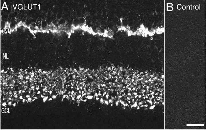
Fig. 1.
VGLUT1 immunoreactivity localizes to the synaptic layers of retina. Photomicrographs of vertical sections through rat retina are shown. A, Confocal fluorescence micrograph of VGLUT1 immunolabeling. VGLUT1 expression is confined to the outer and inner plexiform layers where vesicular glutamate transmission occurs. In the OPL, there is intense VGLUT1 immunolabeling. Labeling is absent from the ONL. Discrete and punctate immunostaining was observed across the full extent of the IPL, with larger puncta observed in the inner portion of the IPL near the GCL. B, There was no immunostaining observed when primary antibody was preabsorbed with VGLUT1 fusion protein. Scale bar, 15 μm.

