
Fig. 3.
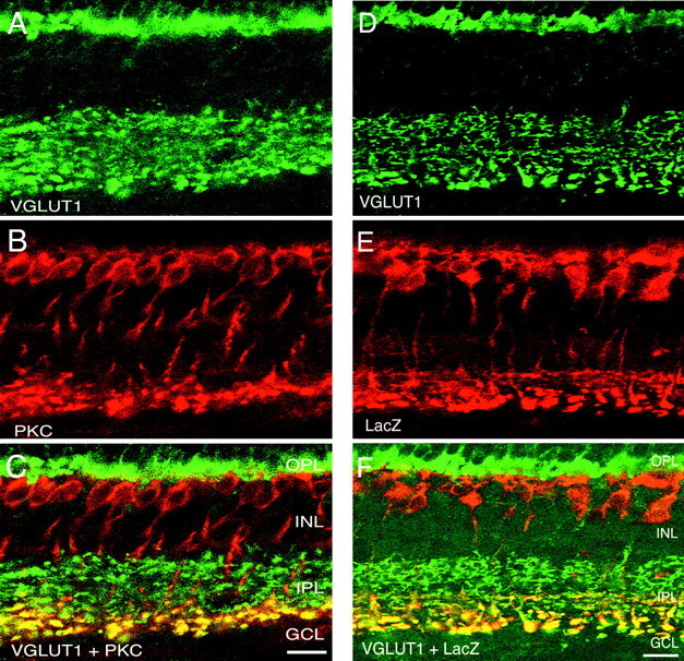
VGLUT1 is expressed in both rod and cone ON bipolars. Confocal fluorescent micrographs of a vertical section of rat retina are shown. A, VGLUT1; B, PKC;C, overlay of VGLUT1 and PKC. Yellowindicates colocalization. VGLUT1 and PKC immunoreactivities overlap in punctate structures located at the inner region of the IPL, consistent with expression of VGLUT1 in rod bipolar cell terminals. Staining was performed in retina from mGluR6+/− transgenic mouse in which LacZ is generated in the ON bipolar cells. D, VGLUT1 staining; E, LacZ antibody staining;F, overlay revealing extensive overlap in the inner half of the IPL, indicating that VGLUT1 is expressed in both rod and cone ON bipolars. Scale bar, 15 μm.