
Fig. 3.
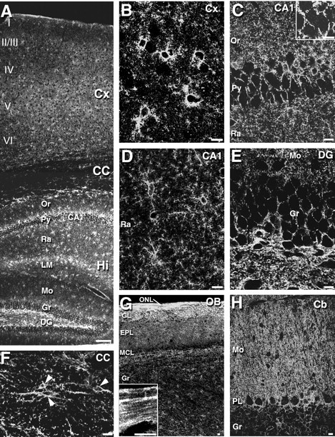
Immunofluorescence for ASCT1 in the adult mouse brain. A, An overview of ASCT1 immunostaining in the cerebral cortex (Cx), corpus callosum (CC), and hippocampus (Hi).B–F, Enlarged views of the cerebral cortex (B), hippocampal CA1 region (C, D), dentate gyrus (E), and corpus callosum (F). The inset inC shows the presence of low immunofluorescent puncta in pyramidal cell perikarya by raising the gain level of the confocal microscope. Arrowheads in F indicate ASCT1-positive cell bodies in the corpus callosum. G, Olfactory bulb. The inset in G is an enlarged image from the olfactory nerve layer (ONL).H, Cerebellar cortex. EPL, External plexiform layer; GL, glomerular layer;LM, stratum lacunosum-moleculare; MCL, mitral cell layer; Or, stratum oriens;I–VI, laminas I–VI of the cerebral cortex. See other abbreviations in the legend to Figure 1. Scale bars: A, 100 μm; B–H, 10 μm.