
Fig. 4.
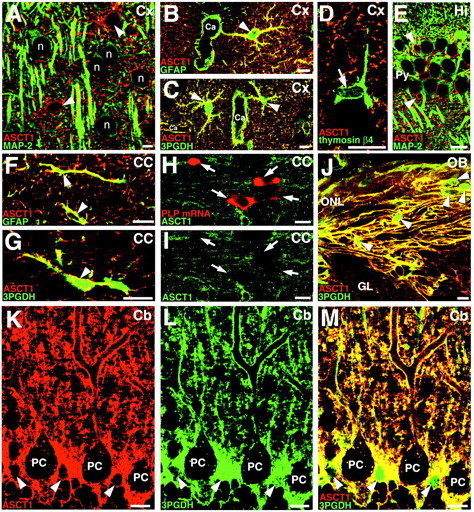
Double staining for ASCT1 and various cellular markers in the adult cerebral cortex (A–D), hippocampus (E), corpus callosum (F–I), olfactory bulb (J), and cerebellar cortex (K–M). In allpanels, ASCT1-immunostained cells are indicated byarrowheads. A, Double immunofluorescence for ASCT1 (red) and MAP-2 (green) in the cortex. ASCT1 is not detected in MAP-2-positive neuronal cell bodies (n) or dendrites. B, ASCT1 (red) and GFAP (green) in the cortex. ASCT1 is detected in GFAP-positive astrocytes (arrowhead), whose processes often surround capillaries (Ca). C, Extensive costaining of ASCT1 (red) and 3PGDH (green) in the cortex. This image is quite similar to B.D, ASCT1 (red) and thymosin β4 (green). ASCT1 is not detected in thymosin-positive microglia (arrow). E, ASCT1 (red) and MAP-2 (green) in the hippocampus. F, Codistribution of ASCT1 (red) and GFAP (green) in callosal astrocytes. G, Codistribution of ASCT1 (red) and 3PGDH (green) in callosal astrocytes. H, I, Double staining for ASCT1 protein (green) and PLP mRNA (red, arrows) in the corpus callosum. ASCT1 expression is lacking in PLP mRNA-expressing oligodendrocytes. J, Extensive costaining of ASCT1 (red) and 3PGDH (green) in the olfactory nerve layer. The interior of olfactory ensheathing glia (arrowheads) is preferentially labeled for 3PGDH, reflecting its cytosolic distribution. K–M, Double immunofluorescence for ASCT1 (red) and 3PGDH (green) in the cerebellar cortex. Cell bodies (arrowheads) and radial fibers of Bergmann glia are costained for both. Note the low particulate immunofluorescence for ASCT1 in the interior of Purkinje cells (PC). Scale bars, 10 μm.